رادیوگرافی پانورامیک
Panoramic
این تصویر برداری اغلب به عنوان یک تصویر ارزیابی اولیه مورد استفاده قرار میگیرد. که میتواند یک دید کلی را تأمین نماید یا در تعیین نیاز به انواع دیگر تصویربرداری ها کمک کند. این رادیوگرافی برای بیمارانی که روند تصویر داخل دهانی را تحمل نمیکنند مفید میباشد
توموگرافی سه بعدی
Cone Beam CT
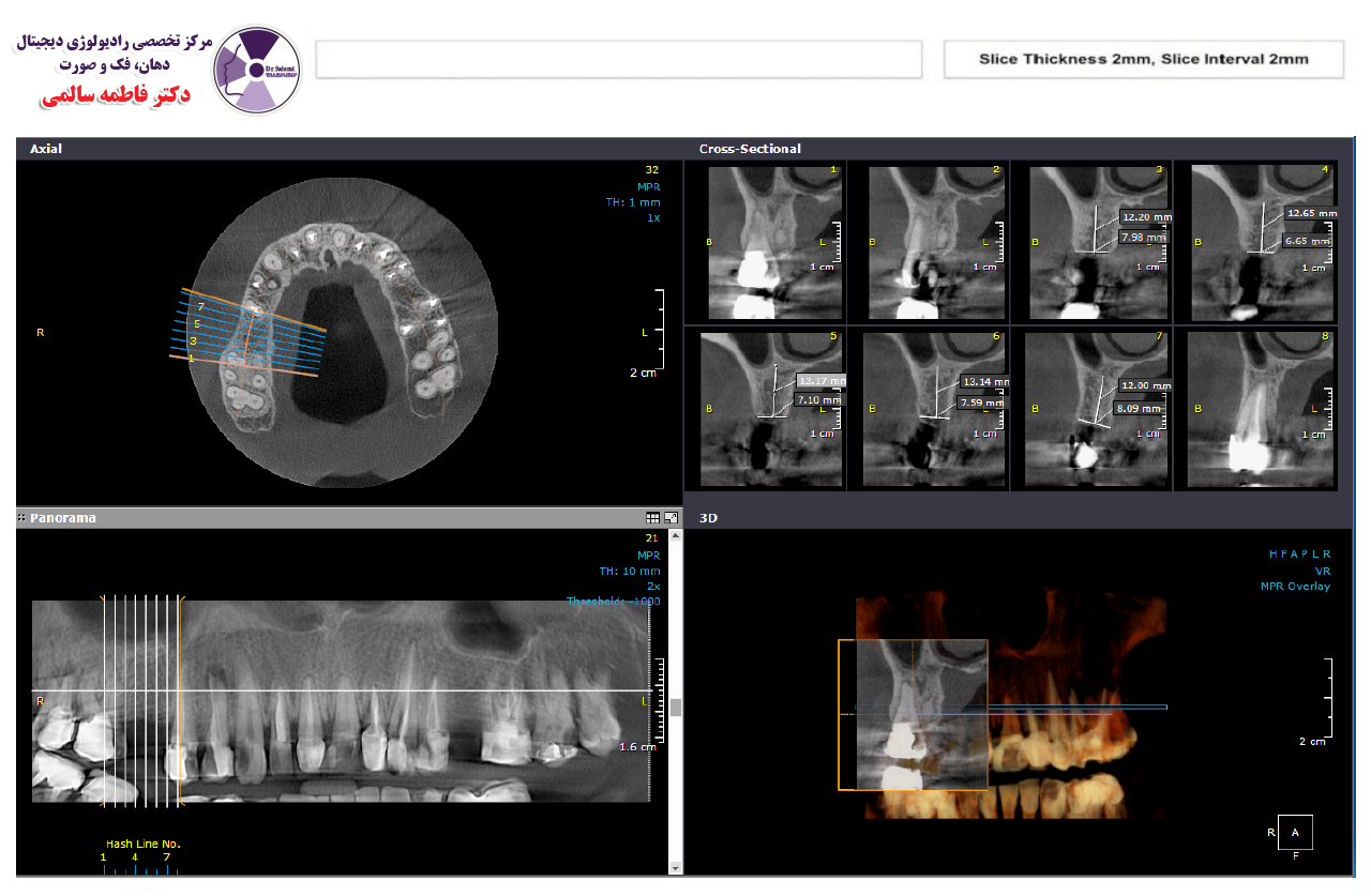
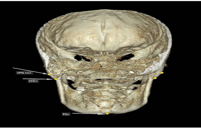
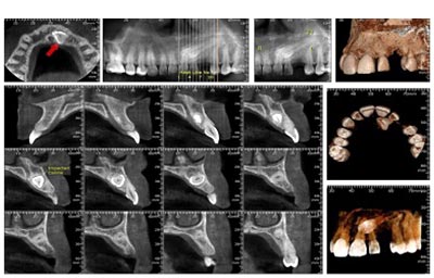
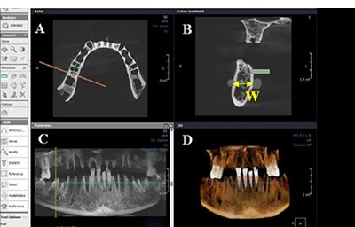
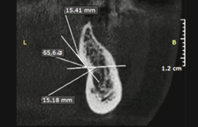
این روش تصویربرداری تصاویر سه بعدی از دندانها، فکین و صورت را در اختیار دندانپزشک قرار می دهد. زمان اسکن کوتاه تر و دوز تابشی بسیار کمتر از CT پزشکی دارد. در این مرکز CBCT با بالاترین دقت و در دو فیلد تابشی متفاوت تهیه می گردد.
رادیوگرافی بایت وینگ
Bitewing
تصاویر بایت وینگ تاج دندان های فک بالا و فک پائین و لبه استخوان فک را نمایش می دهد. تصاویر بایت وینگ برای تشخیص مراحل اولیه پوسیدگی های اینترپروگزیمال و پیش از اینکه از نظر بالینی مشاهده شوند، ارزشمند می باشند.
رادیو گرافی پری اپیکال
Periapical
پرکاربردترین رادیوگرافی برای دنداپزشک میباشد و به تشخیص پوسیدگی و ضایعات التهابی اطراف ریشه کمک می کند. در این مرکز، رادیوگرافی پری اپیکال با روش دیجیتال و با استفاده از نگهدارندههای سنسور استریل شده به تکنیک موازی تهیه میشود. روش موازی کمترین دیستورشن (بدشکلی) را در تصویر ایجاد میکند.
رادیوگرافی لترال سفالومتری
Lateral Cephalometry
رادیوگرافی لترال سفالومتری معمولاً برای ارزیابی و طرح درمان ارتودنسی تهیه میگردد. این رادیوگرافی یک تصویر نیم رخ از استخوانهای صورت ارائه میدهد و در تشخیص ناهنجاریهای دندانی - اسکلتی قبل و حین درمان ارتودنسی کاربرد دارد.
رادیو گرافی اکلوزال
Occlusal
این رادیوگرافی منطقه وسیعی از قوس دندانی یک فک را نمایش میدهد، با سنسور بزرگ تر و عمود بر رادیوگرافی پری اپیکال تهیه میشود و در بررسی موقعیت دندانهای نهفته و سنگهای غده بزاقی کاربرد دارد.
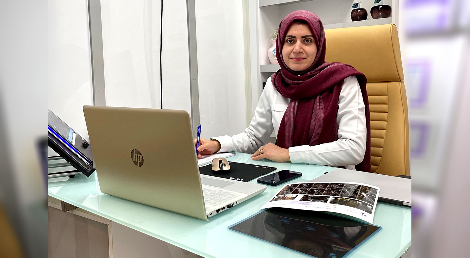
دکتر فاطمه سالمی
دکتر فاطمه سالمی تحصیلات دندانپزشکی خود را در دانشکده دندانپزشکی همدان به پایان رسانده و رشته تخصصی رادیولوژی دهان، فک و صورت را در دانشکده دندانپزشکی اصفهان گذرانده و بورد تخصصی خود را در سال 88 دریافت نموده است. ایشان هم اکنون بعنوان عضو هیئت علمی در دانشکده دندانپزشکی همدان در زمینه های پژوهشی و آموزشی و اجرایی مشغول به فعالیت می باشد و چندین مقاله در مجلات معتبر داخلی و خارجی منتشر کرده است.


