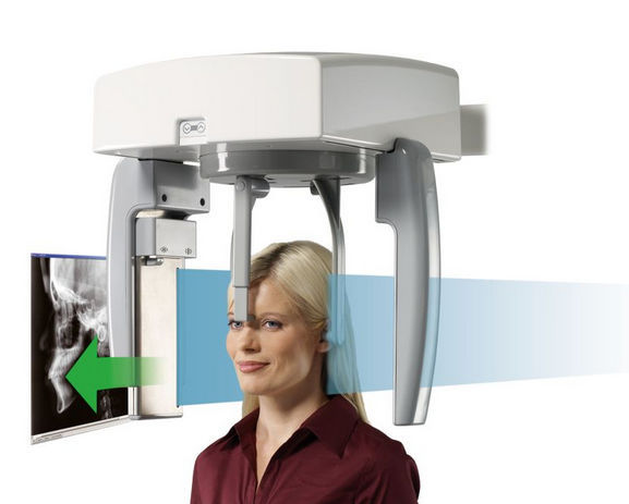
In this radiograph, the patient's teeth are in a closed position and the patient's head is adjusted by two clamps (cephalostat) inside the patient's ears so that the eyes look at the opposite wall. The use of cephaloacetate helps to maintain a stable connection between the head and the image and x-ray receiver. Anatomical points of dental skeleton and soft tissue are defined by lines, angles and distances. At the beginning of the treatment, the measurements are usually compared with certain standards, while during the treatment, the measurements are compared with the previous measurements from the previous cephalometric radiographs of the patient in order to check the growth and development of the patient as well as the treatment process. Lateral cephalometric radiography is usually prepared for evaluation and orthodontic treatment plan, and it is useful for examining the relationship of oral structures. This radiograph provides a profile image of the facial bones in a standard position. In a radiograph, the patient's teeth are in a closed position and the patient's head is held by two clamps (cephalostats) inside the patient's ears so that the eyes are looking at the opposite wall. The use of cephaloacetate helps to maintain a stable connection between the head and the image and x-ray receiver. The anatomical points of the dental skeleton and soft tissue are marked by lines, angles and distances. At the beginning of the treatment, the measurements are usually compared with certain standards. while during the treatment, the measurements are compared with the previous measurements from the previous cephalometric radiographs of the patient in order to check the growth and development of the patient as well as the treatment process.


