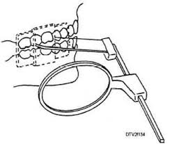
The concept of the parallel technique is that the x-ray receiver is placed parallel to the longitudinal axis of the teeth and the beam is irradiated perpendicular to the teeth and the image receiver. In this method, the distortion or deformity of the image is minimized and it shows the teeth and the supporting bone in their true anatomical relationship. Not long ago or even today, in some centers, periapical radiography was prepared using a semi-automatic method, where the patient had to keep the film in his mouth without shaking or moving it. The advantages of the parallel method compared to the half method:
1- Most of the patients are not able to do this due to stress or pain, also the elderly cannot do this correctly.
2-Also, it is difficult for non-experienced experts to set an angle to perform the graph and increases the possibility of repeating the graph
3- The parallel method depicts the length of the tooth closer to reality.
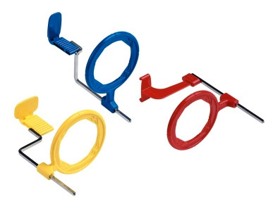
The method of preparing radiographs with the parallel technique: it is done with XCP (Extension cone paralleling) intraoral retainers. These retainers include: plastic bite block (bite block), guide rod and an aiming ring. These components must be correctly be installed, that is, the byte block and the film should be in the center of the ring.
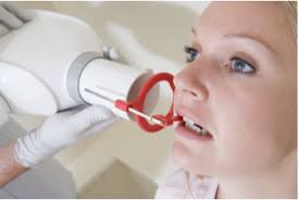
There are three types of retainers, including anterior-posterior and bitewing, each of which is made in the same color for the convenience of the technician.
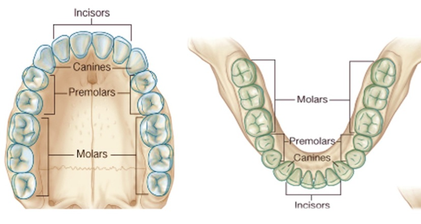
To perform periapical photography of all teeth, the oral cavity is divided into 4 areas, which are examined with 14 films.
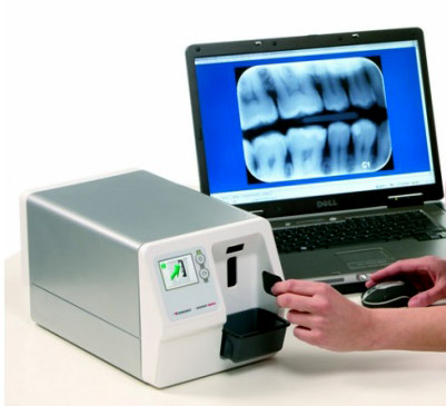
In this center, radiographs are done digitally using a phosphor plate (PSP) sensor. Digital radiography is a method in which an X-ray sensitive sensor is placed in the patient's mouth instead of radiographic film. Dental digital imaging is performed by two systems of RVG sensors and phosphor plate scanners.
Phosphor plate sensors:
Phosphor plate sensors : are wireless and their thickness is even less than radiographic films. The working method of phosphor plates is very similar to the imaging method with film. After the image is taken, the sensor is removed from the patient's mouth and scanned by a scanner on the monitor.
The computer is displayed Advantages of digital radiography :
Quick view of the photo: This advantage is very desirable during most treatments and especially during the process of implant placement, root canal treatment and also patient education.
Save information: Easy storage and electronic playback of digital photos allows better communication with other dentists for easy comparison and follow-up.
Reduction of radiation: Based on the type of radiography used, radiation to the patient is greatly reduced compared to old methods.
Removal of chemicals and dark room: The maintenance of developing solutions and film proofing and darkroom conditions determine why radiographic images with film can be removed and converted to digital radiography.
